Sie sind hier: Skip Navigation LinksInstitut für Anatomie Forschung Finanzierte Projekte Tumorschnittkulturen

Tumorschnittkulturen

(EN: Tumor-slice-culture/tumorslices)

​​​Mausmodelle und primäre Zellkultur sind gängige Modelle in der Tumorforschung und bilden die Grundlage für die klinische Therapie. Die mit diesen Methoden erzielten Ergebnisse sind jedoch aufgrund interspeziesischer Unterschiede und interzellulärer Mechanismen nur schwer auf den menschlichen Organismus übertragbar. 

Darüber hinaus erfordern die großen Auswirkungen von Heterogenität, Immunologie und Stroma-assoziierten Modulationen in der Tumorbiologie neue Strategien zur Untersuchung des komplexen Gewebegleichgewichts. Organotypische Schnittkulturen menschlicher Krebserkrankungen sind geeignet, ein besseres Verständnis bestimmter interzellulärer Prozesse zu erlangen und können so die Übersetzungsschritte von der zellulären und mausbasierten Forschung bis zur klinischen Umsetzung verkürzen. 

Der Schwerpunkt dieses vom BMBF geförderten Projekts liegt auf der Untersuchung tumorbedingter Resistenzmechanismen und dem Aufbau eines patientenorientierten Tools zur Vorhersage der Anfälligkeit für bestimmte Therapien.​

​​Kein Porträtfoto vorhanden ​Prof.in Dr.in Sonja Kallendrusch

Publikationen

E-Mail: sonja.kallendrusch@medizin.uni-leipzig.de
Porträt eines lächelnden älteren Mannes mit randloser runder Brille ​Prof. Dr. Ingo Bechmann

Publikationen

E-Mail: ingo.bechmann​@medizin.uni-leipzig.de
Telefon: 0341 - 97 22001
​​

PROJEK​T 1

Schnittkulturen für eine personalisierte Suszeptibiliätsüberprüfung​

In Zusammenarbeit mit Prof. Florian Lordick, MD Justus Körfer, MD Sebastian Pill, MD Laura Hennig und MD Rasmus Sönnichsen

Adenokarzinome des gastrointestinalen Traktes sind teilweise sehr aggressive und sehr heterogene Tumorentitäten. Eine hohe Inzidenz und die zum Teil sehr schlechte Prognose, da das Ansprechen auf Chemotherapie oft ausbleibt, bedingen die Notwendigkeit ein personalisiertes Therapieverfahren zu ermitteln. Tumorproben aus OP Präparaten und Biopsien werden in Schnittkulturen angelegt und mit unterschiedlichen Chemotherapeutika behandelt. Das Ziel ist es, nach erfolgreicher Etablierung der Methodik, dies klinisch zu korrelieren um somit geeignete Therapiekonzepte für den individuellen Patienten entwickeln zu können.​

Kallendr_P1.jpg

Figur 1: Colon Cancer AE1 3 (rot) Ki67 (grün) DAPI Blau 40x

PROJEKT 2

Schnittkulturen sind immunkompetent und eignen sich zur Testung von Immunmodulatoren

In Zusammenarbeit mit Dr. Sebastian Krämer, Prof. Florian Lordick, Prof. Uwe Eichfeld und David Junk, MD student

Tumore der Lunge stellen eine der häufigsten Todesursachen im Zusammenhang mit Tumoren dar. Neuere Medikamente, monoklonale Antikörper (Nivolumab, Pembrolizumab) sind sogenannte Immunomodulatoren und aktivieren die T-Lymphozyten- Tumor- Interaktion. Nivolumab ist ein zugelassenes Medikament, welches die Interaktionszeit von T-Zellen und Tumorzellen erhöht. Diese verlängerte Kontaktzeit kann für ein erkennen der Tumorzelle sorgen und eine entsprechende Immunantwort ausgelösen. Schnittkulturen von Lungenkarzinomen stellen eine geeignete Methode dar um diese Immunomodulatoren im Gewebeverband zu untersuchen und auf ein individuelles Ansprechen zu testen.

Kallendr_P2.jpg

Figur 2: DiV3 20x DAPI (blau) Glut1 (rot) Ki67 (grün) Lungenkrebs

PROJEKT 3​

Humane organotypische Schnittkulturen aus Glioblastomgewebe als Testsystem zur Auf-klärung molekularer Mechanismen der Therapieresistenz und Tumorausbreitung

In Zusammenarbeit mit Prof. Frank Gaunitz und Prof. Jürgen Meixensberger und Dr.in Susann Hähnel

Der Fokus dieses Projekts liegt auf der Identifikation von Resistenzmechanismen von Glioblastoma multiforme gegen die heutige Standardtherapie, bestehend aus Chemotherapie und Bestrahlung, und der Ausbreitung der Tumorzellen in das umliegende gesunde Gewebe. Zudem soll in einer prospektiven Studie die Suszeptibilität in der Slice-Kultur mit individuellen Verlaufsparametern verglichen werden. Durch Sequenzierung des Transkriptoms von Glioblastomgewebe sollen Merkmale identifiziert werden, die für Diagnostik, Therapie und Prognose von Bedeutung sind.​​

Kallendr_P3.jpg

Figur 3: DiV7 10x HE Glioblastoma multiforme

PROJEKT 4

Gentransduktion von humanen Glioblastoma Spheroidkulturen mit Hilfe von Adeno-assoziierten viralen (AAV) Vektoren​

In Zusammenarbeit mit MD Jule Ußmann, PD. Ulrich Hacker, Prof. Frank Gaunitz, Prof. Jürgen Meixensberger und Prof.in Hildegard Büning

​Glioblastoma multiforme ist eine maligne Tumorentität mit einer sehr schlechten Prognose. Tumorformende, stammzellartige Zellen überleben weitestgehend die übliche Temozolomid-, sowie die Strahlenbehandlung und stellen somit eine Population dar, die gentechnisch für eine rekombinante Adeno-assoziierte virale Vektor (rAAV) Manipulation geeignet sind. Es soll untersucht werden, ob der in Zelllinien etablierte Serotyp auch in primären Spheroiden eine Transduktion bewirkt und wie tief er die Spheroidkulturen durchdringt um anschliessend gentechnische Manipulationen durch Designer-Endonukleasen (Gen EFNB2, EphrinB2) in Spheroidkulturen und Schnittkulturen durchzuführen. Hiermit soll die Anwendbarkeit und die Interaktionen von AAVs im intakten Gewebe untersucht werden.​​

Kallendr_P4.jpg​​​Figur 4: AAV GFP PI (rot) Hoechst (blau) 10x


IN KOOPERATION MIT UCCL​

UCCL.jpg
Klicken Sie auf das Bild, um auf die Website des UCCL zu gelangen

Ehemalige Labormitarbeiter: Felicitas Merz PhD | Magdalena Gerlach MD | Justus Körfer MD | Nikolas Schopow MD​​​

englischsprachige Projektbeschreibungen

Mouse models and primary cell culture are common models in the field of tumor research and build the foundation for clinical therapy. The results gained by those methods are however poorly adaptable to the human organism due to inter-species differences and intercellular mechanisms. Further, the high impact of heterogeneity, immunology and stroma associated modulations in tumor biology require new strategies to investigate complex tissue equilibration. Organotypic slice cultures of human cancers are suitable to obtain an enhanced understanding of certain inter-cellular processes and thus can reduce the translational steps from cellular and mouse-based research towards clinical implementation.

The focus of this BMBF-funded project relies on the investigation of tumor-related mechanisms of resistance and to build up a patient-oriented tool to predict susceptibility for certain therapies. ​​


PROJECT 1

Organotypic slice cultures of human cancers of the GI tract

In cooperation with Prof. Florian Lordick, MD Justus Körfer, MD Sebastian Pill, MD Laura Hennig and MD Rasmus Sönnichsen

Poor prognosis and high biologic heterogeneity are main characteristics of colon and gastric cancer. The high rate of patients that do not respond to cytotoxic treatment urge for predictive assays for the individual patient. We aim to standardize the organotypic tumour slice culture system to investigate cytotoxic drug susceptibility prior to patient treatment, which may lead to personalised and more precise treatment strategies.

Kallendr_P1.jpg

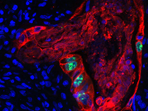
Figure 1: Colon Cancer AE1 3 (red) Ki67 (green) DAPI Blue 40x

PROJECT 2

Organotypic slice cultures of human lung cancer

In cooperation with Dr. Sebastian Krämer, Prof. Florian Lordick, Prof. Uwe Eichfeld, MD David Junk

Lung cancer belongs to the most malignant tumor entities and although therapeutic advances could be made in surgery techniques and cytostatic treatments, the prognosis remains poor. Novel therapies, targeting the immune system, promise beneficial effects. Still, there are predictive assays needed that prevent ineffective treatment, as not all patients respond equally to applied therapies. A standardized organotypic tumor slice culture system offers the opportunity to study the individual tumor towards its susceptibility to different drugs.

The leading hypothesis of this project is that nutrition-induced cerebral accumulation of fatty acids, related bioactive lipids and their metabolites can promote microglial senescence and loss, e.g. by causing vascular stiffness and/or peroxysomal

Kallendr_P2.jpg

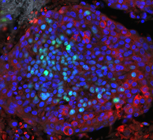
Figure 2: DiV3 20x DAPI (blue) Glut1 (red) Ki67 (green) Lung Cancer

PROJECT 3

Organotypic slice cultures of human glioblastoma multiforme tissue

In cooperation with Prof. Frank Gaunitz and Prof. Jürgen Meixensberger, Dr. Susann Hähnel

Glioblastoma multiforme often and quickly develops resistance towards standard treatment, consisting of chemotherapy and radiation. Organotypic slice cultures of human glioblastoma multiforme tissue can be cultivated and treated with the clinical standard protocol. The project aims to correlate the response of patient-derived slice cultures with the prospective follow up of the corresponding patient. Furthermore, the underlying mechanisms which allow the tumor to escape therapy and to spread even faster into healthy brain tissue are studied by deep sequencing data of patient-derived glioblastoma multiforme tissue.

Kallendr_P3.jpg

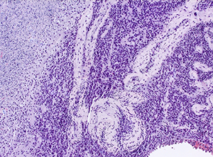
Figure 3: DiV7 10x HE Glioblastoma multiforme

PROJECT 4

Adeno-associated Viral Vector transduction in glioblastoma multiforme

In cooperation with PD. Ulrich Hacker, Prof. Frank Gaunitz, Prof. Jürgen Meixensberger und Prof. Hildegard Büning, MD Jule Ußmann

Glioblastoma multiforme represents the most malignant brain tumor with poor prognosis and high recurrence rates. Recurrence is believed to be induced by stem cell like cancer stem cells which are resistant to standard therapy. Primary neurosphere cultures display the stem cell like tumor cells, providing an exellent setup to investigate resistance mechanisms and novel stategies to combat these tumor-inducing cells. Adeno-associated virus (AAV) vectors have gained an increasing attention as gene therapy vectors for several diseases. The first clinically approved AAV-based gene therapy in the Western world is Glibera® demonstrating that this approach can successfully and safely be applied. The project studies AAV targeting vectors in primary human spheroid cultures and organoid tissue cultures of glioblastoma multiforme tissue.

Kallendr_P4.jpg
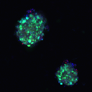
Figure 4: AAV GFP PI (red) Hoechst (blue) 10x

PROJECT 5

Methodological Investigation of organotypic slice cultures

In cooperation with Vera Blaschke, master student

Monolayer cell culture models are lacking extracellular cues that can affect cellular phenotype and therewith display different results compared to three-dimensional cell culture. Recent studies have shown that three-dimensional cell and tissue culture are valuable tools for drug testing, cellular investigations but also mean to become beneficial in personalized medicine. Though application of standard methods like PCR, WB or histology is applicable, FACS analysis however needs to be established as tissue dissociation is crucial for cellular expression profiles. FACS analysis is time-saving and allows investigating changes of various cell populations or cellular changes within one tumor sample. The present master thesis aims to define a tissue dissociation protocol well adapted for long term tissue culture. Organotypic tumor slice cultures will be studied for immunological populations such as microglia/macrophages and T-cell populations.​​

Kallendr_P5.jpg


​​​​​FINANZIERUNG

Liebigstr. 13, Haus A
04103 Leipzig
Telefon:
0341 - 97 22000
Map